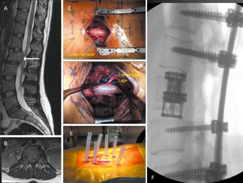
Figure 6.

A 32‐year‐old woman presented with mid back pain and leg weakness over 3 weeks. (A) and (B) MRI images showing solitary L2 lesion causing circumferential cauda equina compression. (C) Intraoperative photograph showing midline incision with circumferential decompression and vertebrectomy. Decompressed L3 nerve roots are displayed (arrow). (D) Intraoperative photograph showing expandable cage (arrow) assisted vertebral body reconstruction. (E) Intraoperative photograph showing midline wound closure and percutaneous fixation two levels above and below the vertebrectomy. (F) Post‐operative radiograph.
