
Figure 2.
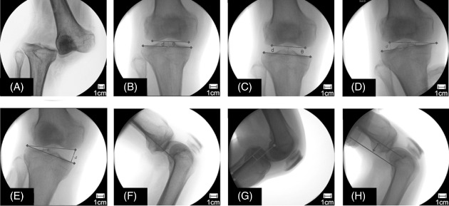
Preoperative evaluation of the ligamentous laxity under different stress conditions. (A) Radiological appearance of a dislocated knee in the emergency room. (B) The reduced knee showing a normal articular match (distance of the joint gap [d] = 7.5 mm, angle of the articular surface [θ] = 0°). (C) An axial pulling view showing equally widened medial and lateral joint spaces (d = 13.8 mm, θ = 0°). (D) A varus stress radiograph showing a Grade II injury of the LCL (d = 13.2 mm, θ = 8.5°). (E) A valgus stress radiograph showing a Grade III injury of the MCL (d = 21.3 mm, θ = 14.2°). (F) A lateral radiograph of the knee maintained in the neutral position. (G) Posterior movement of 8.8 mm of the tibia to the femur, indicating a torn PCL. (H) Anterior movement of 19.2 mm of the tibia on the femur, indicating a torn ACL.