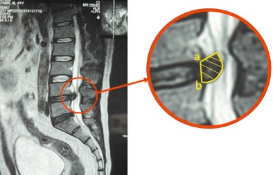
Figure 1.

T 2 weighted sagittal view MR image showing how the resorption rate is calculated. The internal boundary of the protrusion is the connecting line between the posterior inferior margin of the upper centrum and the posterior superior margin of the lower centrum. The external boundary is the protrusion edge. A proficient MRI operator can determine the area of the protrusion, as shown in the right panel. The volume of the protrusion (mm3) = (inter‐section spacing + section thickness) (mm) × Σ area of the protrusion in each section (mm2). The resorption rate = (volume of protrusion before treatment—volume of protrusion after treatment/volume of protrusion before treatment) ×100(%).
