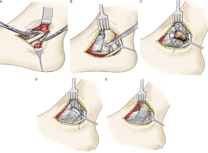
Figure 3.

Diagram of the surgical procedure: (A) The posterior tibial tendon is exposed. (B) Approximately 5 mm from the anterior colliculus of the medial malleolus, a transverse incision is made in the deep layer of the posterior tibial tendon sheath and superficial layer of the deltoid ligament. (C) A Smith‐Nephew 3.5 mm suture anchor is placed in an anterior‐to‐posterior direction. (D) The distal end of the superficial layer of the deltoid ligament is sutured and fixed onto the bone structure of the anterior colliculus of the medial malleolus. (E) The periosteum and proximal end of the superficial deltoid ligament layers are overlapped and sutured onto the distal severed end and reinforced with suture anchors.
