
Figure 4.
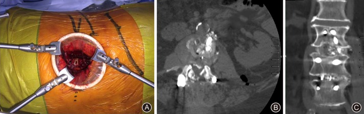
(A) Intraoperative view of the oblique corridor, with the psoas and retroperitoneum retracted using a Synframe. (B) Follow‐up computed tomography (CT) (transverse) of interbody cage. Arrow demonstrates oblique corridor used for revision of non‐union after previous fusion surgery. (C) Follow‐up CT (coronal) demonstrating appropriately positioned new revision cage inserted using an oblique trajectory.