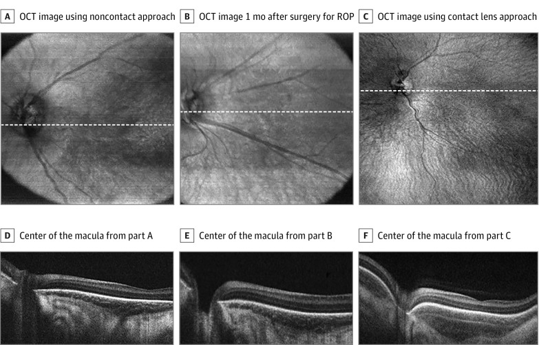
Figure 1. Optical Coherence Tomography (OCT) in Children in the Neonatal Intensive Care Unit With Retinopathy of Prematurity (ROP) Using Handheld Prototype Swept-Source OCT.
A, En face OCT image of a neonate obtained in 2 seconds during routine screening using a noncontact approach. B, En face OCT image of a neonate obtained in 2 seconds during routine screening using a noncontact approach 1 month after laser surgery for type 1 ROP. C, En face OCT image of a neonate obtained obtained in 2 seconds during routine screening using contact lens approach. D, Horizontal line scan through the center of the macula of the neonate from part A. E, Horizontal line scan through the center of the macula of the neonate from part B. F, Horizontal line scan through the center of the macula of the neonate from part C. White dashed lines in A represent corresponding line scans in D, white dashed lines in B represent corresponding line scans in E, and white dashed lines in C represent corresponding line scans in F.

