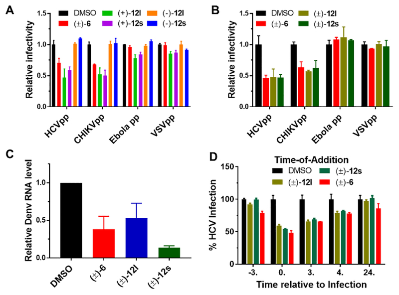
Figure 4.
Aglaroxin C (6) and its analogues (121 & 12s) inhibit viral entry. (A) Huh7.5.1 cells were infected by indicated pseudovirus in the presence of compounds (1 μM) for 3 h. After an additional 48 h infection, cells were lysed for luciferase assay. Relative infectivity was calculated by normalizing against the values obtained from cells treated with DMSO (arbitrarily set to 1.0) (mean ± SD, *p < 0.05). (B) Same as in a except (±)−121 and (±)−12s were added at 2 μM. (C) Huh7.5.1 cells were infected by Dengue virus (serotype 2, Thailand 16681 strain, MOI 0.1) treated with 2 μM compounds for 3 h. 48 h post-infection, RNA was isolated for quantitative RT-PCR analysis. Viral RNA levels were normalized against GAPDH levels. Data presented are representatives of two independent experiments. (D) (±)−6, (±)−12l, and (±)−12s inhibited HCV infection when added together with the virus. Details on this time-of-addition experiment can be found in Supporting Information.

