
Figure 2.
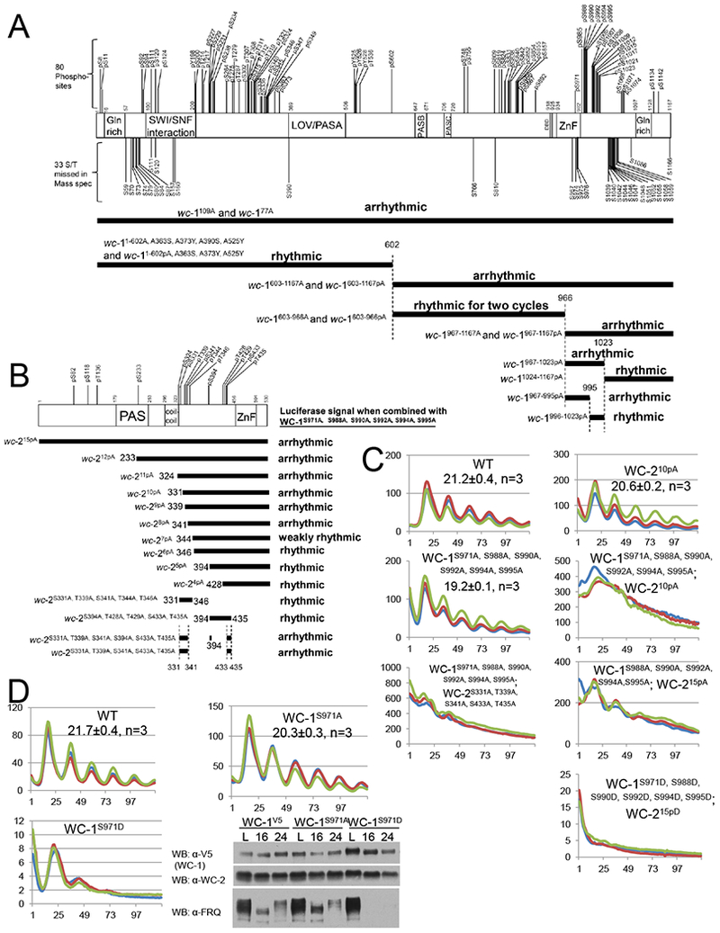
Identification of key phosphosites on WC-1 and WC-2 required for the circadian feedback loop closure. Schematic of WC-1 (A upper) and WC-2 (B upper) showing the position of phosphorylated residues as in Figure 1BC. Below these is the strategy used to identify the phosphorylation events on WC-1 (A lower) and WC-2 (B lower) essential for rhythmicity. Each horizontal bar represents a wc-1 (A) or wc-2 (B) mutant with phosphosites falling in the region of the bar mutated to Ala altogether. In the presence of wc-215pA (A) or wc-1S971A, S988A, S990A, S992A, S994A, S995A (B) respectively, the circadian rhythms of the wc-1 (A) or wc-2 (B) phosphomutants were measured by frq C-box-luc ; phenotypes (Table S4) were as noted. C WCC activity in WT and wcc mutants (y-axis) as measured by frq C-box-luc bioluminescence. D Phosphorylation of WC-1 S971 plays a key role in repressing the circadian WCC activity. WCC activity in strains of the noted genotypes was monitored by frq C-box-luc; note scale bars. Right panel: Western blot showing FRQ, WC-1, and WC-2 levels in WT (wc-1V5), wc-1S971A, and wc-1S971D in light and at 16 and 24 hrs in darkness.