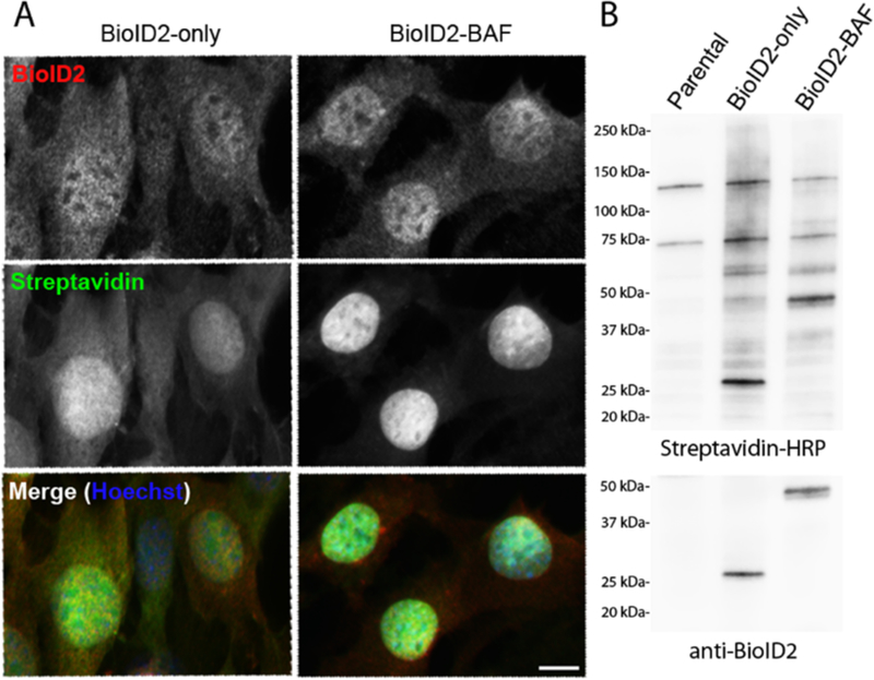
Figure 2.

Representation of stable BioID-fusion protein expression in cells. NIH3T3 cells stably expressing BioID2-only or a BioID-fusion protein of interest (BioID2-BAF) were analyzed for fusion protein expression, localization and biotinylation by fluorescence microscopy and immunoblot. A) Using florescent microscopy, fusion proteins were detected by anti-BioID2 antibody (red) and biotinylation was detected by streptavidin-488 Alexa Fluor (green). Scale bar = 10 µm. B) Biotinylation efficiency and fusion protein expression in whole cell lysate were also assessed by western blot, using streptavidin-HRP and anti-BioID2 antibody.
