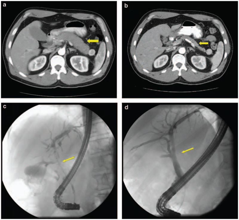
Figure 2.
Contrast-enhanced computed tomography in a patient with autoimmune pancreatitis (a) before initiating steroid treatment showing diffuse (sausage-shaped) pancreatic enlargement and (b) after 6 weeks of steroid treatment showing a decrease in size of the pancreas. Cholangiogram in a patient showing (c) extensive hilar stricturing before treatment and (d) after treatment showing resolution of strictures; 165×152 mm (96 × 96 d.p.i.).

