
Figure 3. Structures of initiation factors TFAM and TFB2M.
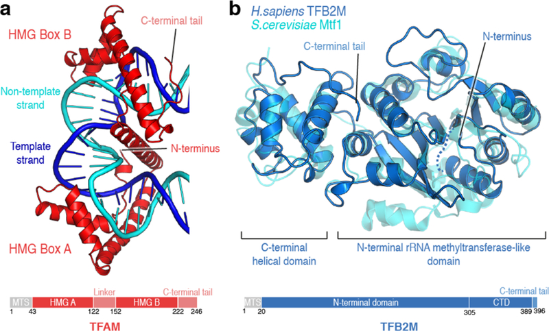
(a) Ribbon depiction of human TFAM bound to the LSP promoter sequence (PDB ID: 3TMM) colored in red. DNA coloring as in Figure 2. Important structural elements are indicated. A schematic depiction of the domain architecture of TFAM is shown below. MTS: Mitochondrial targeting sequence. Each of the HMG box domains of TFAM induces an approximately 90° bend to the duplex DNA by intercalating a hydrophobic residue, resulting in a 180° turn. (b) Ribbon depiction of human TFB2M (PDB ID: 6ERO) colored in marine blue with the structure of the yeast mitochondrial transcription initiation factor Mtf1 (PDB ID: 1I4W) overlaid transparently in cyan. A schematic depiction of the domain architecture of TFB2M is shown below. MTS: Mitochondrial targeting sequence. The N-terminal part of TFB2M and Mtf1 adopt a rRNA methyltransferase-like fold, while the C-terminal part is a globular, all-helical domain. TFB2M has a flexible tail which is important for its function in transcription.