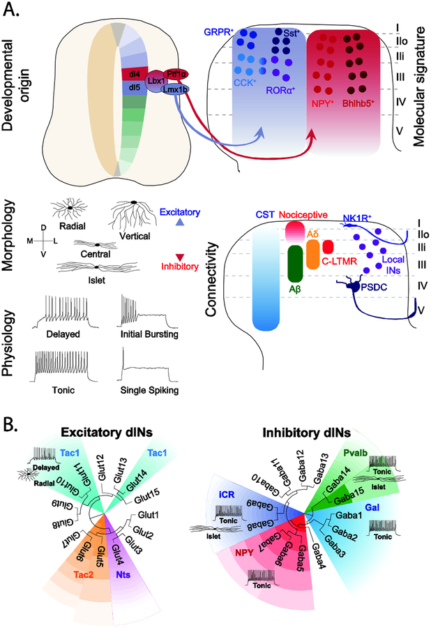
Figure 2. Dorsal horn neuron diversity.
A. Spinal cord cell types have been classified according to their developmental origin, expression of defined molecular markers, morphology, physiology and connectivity. Dorsal horn neurons that process and gate noxious and innocuous cutaneous sensory information arise from Lbx1+ dI4 and dI5 progenitors that are marked by the expression of Lbx1 and express several post-mitotic markers [10]. Dorsal horn neurons can also be classified according to their morphological and electrophysiological properties as exemplified by the classification of two neurochemically distinct neuron types: GRP+ and Tac1+ INs [48]. Neurons in the more superficial laminae, receive little corticospinal (CST) and strong noxious input, whereas neurons within the LTMR-RZ receive a unique mix of Aβ-, Aδ- and C-LTMR and CST input [35••]. Lamina position is also a determinant of identity, with NK1R+ projection neurons in lamina I contributing to the Spinothalamic Tract [52], and neurons within laminae III/IV being part of the Post-Synaptic Dorsal Column (PSDC) [35••].
B. ScRNA-seq analysis of dorsal horn neurons showing the transcriptomic clusters identified in Häring et al. [25••]) overlaid with known neurochemical markers, morphology and physiology [26•,28,30,48–51,53]. Tac1: Tachykinin 1, Tac2: Tachykinin 2, Nts: Neurotensin, iCR: inhibitory Calretinin, NPY: Neuropeptide Y, Pvab, Parvalbumin, Gal: Galanin.

