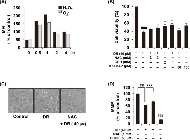
Fig 4. Increased ROS production by DR treatment is related to autophagy.
(A) After A549 cells were treated with DR, the cells were loaded with the hydrogen peroxide sensitive dye H2DCFDA or the superoxide sensitive dye HE at 37°C for 30 min, and ROS generation was then measured using flow cytometry. (B) Cells were treated 40 μM DR for 24 h with or without treatment with NAC, GSH, and MnTBAP. The cell viability was measured by WST assay. (C) The morphological change of cells was observed under the microscope. (D) The cells were incubated with DiOC6(3) for 30 min 37°C in the dark, and analyzed by a flow cytometery. Carbonyl cyanide 3-chlorophenylhydrazone (CCCP; 40 nM) was used as a positive control. Statistical differences were presented p<0.05 (*), p<0.01 (**), and p<0.001 (***) compared with the DR alone; p<0.01 (##) compared with the DMSO control.

