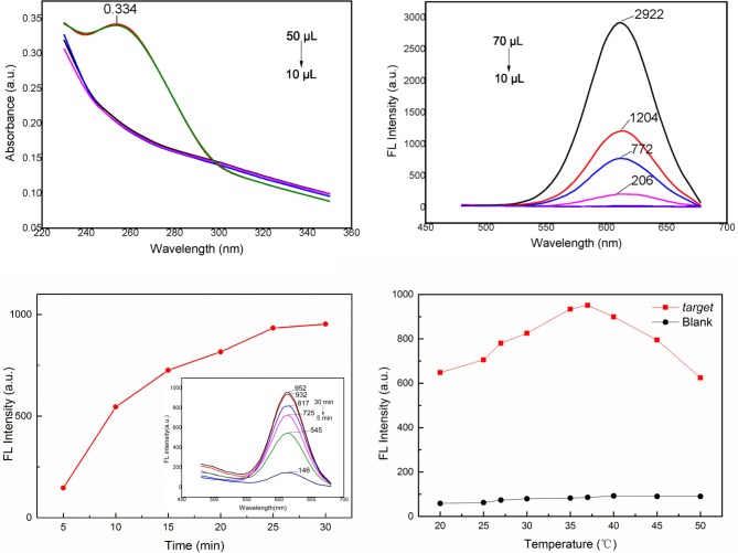
Fig 6.
(a) UV-visible absorption spectrum of 10 μL of 1 mg•mL-1 streptavidin-coated MNPs decorated with 50 μL of 10 nM aptamer. (b) Fluorescence spectra of different concentrations (from 70 μL to 10 μL) of ssDNA2@CdTe QDs of 30 μg·mL-1 ssDNA2@CdTe QDs. (c) Fluorescence spectra of aptamer&QDs-ssDNA2@MNPs after different incubation times with S. Typhimurium. (d) Fluorescence spectra of aptamer&QDs-ssDNA2@MNPs incubated with S. Typhimurium at different incubation temperatures.

