Figure 3.

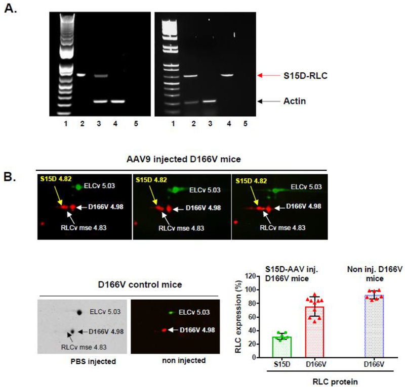
A. PCR detection of AAV9 genome in the ventricles of non-transgenic (NTg) mice (left) and D166V HCM transgenic (Tg) mice (right) 7 days post injection. Left PCR panel: Lane1- DNA ladder, Lane2-vector positive ctr, Lane3-AAV9-S15D-RLC injected NTg mouse ventricle, Lane4-non-injected control NTg ventricle, Lane5-water ctr. Right PCR panel: Lane1- DNA ladder, Lane2-AAV9-S15D-RLC injected D166V ventricle, Lane3-non-injected D166V ventricle, Lane4-vector positive ctr, Lane5-water ctr. Red arrow – diagnostic band for AAV9-S15D-RLC genome; black arrow – actin (internal control). B. 2D-SDS-PAGE detection of human S15D-RLC in myosin from left ventricles of AAV9 injected (at 1.4×10¹¹ vg) D166V (Western blots) vs PBS (Coomassie) or non-injected (Western blot) D166V mice. Expression (E) of S15D was calculated using the formula E = S15D/(S15D+mseRLC+D166V). Average S15D-RLC expression in LV of AAV-injected D166V mice (by 2D-electrophoresis of n=7 samples from 5 animals) was 31±5% (Avg±SD). Average D166V RLC expression in D166V non-injected mice was 93±6% (n=7 samples from 2 animals) and 76±14 (n=10 samples from 6 animals) on AAV-S15D-RLC injected D166V mice.
