
Figure 3. Entinostat suppresses the EWS-FLI1/HDAC3/HSP90 signaling axis in ES cells.
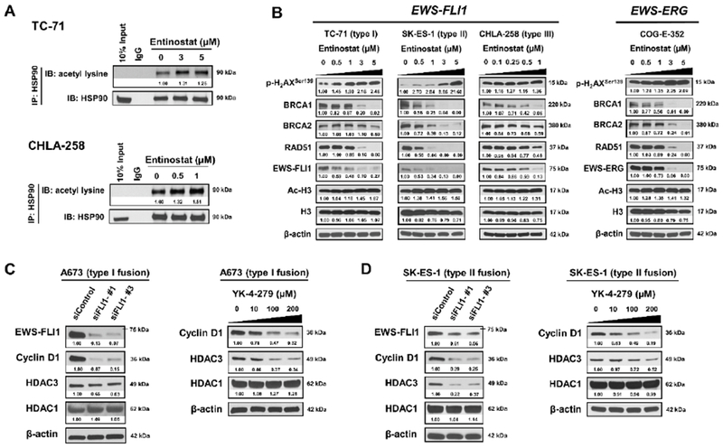
A, Entinostat induced hyperacetylation of HSP90 in both TC-71 and CHLA-258 cells 6 hours post treatment. IgG, immunoglobulin G. B, Representative Western blots showing changes in protein expression in TC-71, SK-ES-1, CHLA-258 and COG-E-352 cells after 48 h incubation with entinostat. Cell lysates were probed with the indicated antibodies. C, Knockdown of EWS-FLI1 leads to the downregulation of HDAC3 expression in A673 cells at 72 h post transfection. Meanwhile, YK-4–279 treatment reduced the expression of HDAC3 dose-dependently in A673 cells at 12 h post treatment. β-actin served as a loading control. D, Knockdown of EWS-FLI1 leads to the downregulation of HDAC3 expression in SK-ES-1 cells at 72 h post transfection. Meanwhile, YK-4–279 treatment reduced the expression of HDAC3 dose-dependently in SK-ES-1 cells at 12 h post treatment. Cyclin D1 is a validated transcriptional target of EWS-FLI1 and served as a positive control. β-actin served as a loading control. The relative intensities of protein bands are shown under the immunoblots after normalization for the levels of β-actin.