
Figure 1.
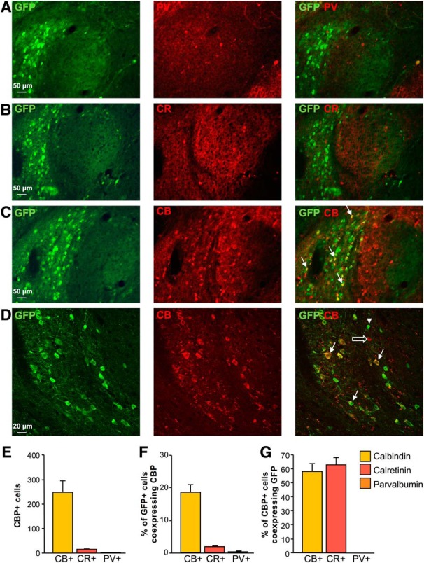
CBP expression in CRF1+ cells in the CeA. Representative images of double immunostaining for GFP and (A) PV (red, GFP = green), (B) CR (red, GFP = green), and (C, D) CB (red, GFP = green) in the CeA of CRF1:GFP mice. Co-expression patterns are visualized in overlaid images in the third column where the arrowhead indicates a single-labeled GFP+ cell, the open arrow indicates a single-labeled CB+ cell, and filled arrows indicate double-labeled cells that co-express GFP and CB. Scale bars = 50 μm (A–C) and 20 μm (D). E, Summary bar graph representing the total number of CBP+ cells counted in the CeA. F, Bar graph representing the proportion of CRF1+ cells co-expressing each CBP. G, Bar graph representing the proportion of CBP+ cells co-expressing CRF1. Data are shown as mean ± SEM. Figure Contributions: H. Sidhu performed the experiments and analyzed the data.