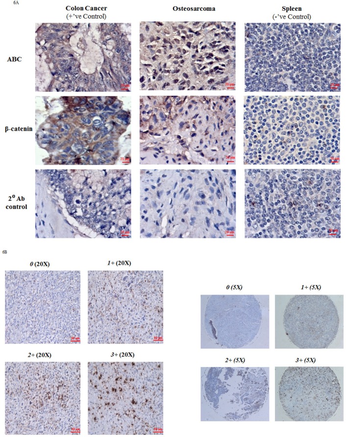
Figure 6. Immunohistochemical analysis of ABC and β-catenin in OS tissue.
A. Figure shows immunoexpression of ABC and β-catenin in OS tissue. Colon cancer tissue served as a positive tissue control and spleen tissue was used as negative tissue control. Immunoexpression data is representative of 6 experiments. B. Representative data of nuclear ABC protein staining in osteosarcoma tissue cores on TMA slide at magnification 20X and 5X (entire core). 0 (no nuclear staining); 1+ (≤30%); 2+ (31-60%); 3+ (>60%).

