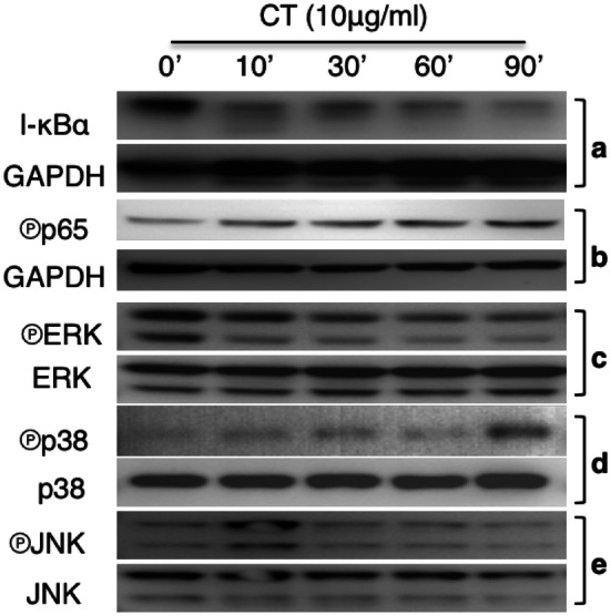
Fig. 3.

CT regulation of NF-κB and MAPK activation. a Mouse bone marrow-derived DCs treated with 10 µg/ml of CT for the indicated time periods were solubilized in 1 × SDS sample buffer at 107/ml. The samples were separated on a 4–12% gradient NuPAGE™ gel, transferred on a piece of Immobilon™ membrane, blocked, and treated with anti-IκBα (a), anti-phospho-p65 (b), anti-phospho-Erks (c), anti-phospho-p38 (d), or anti-phospho-JNK (e). After the images were taken, the membranes were stripped and re-probed with anti-GAPDH, anti-Erks, anti-p38, and anti-JNK, respectively
