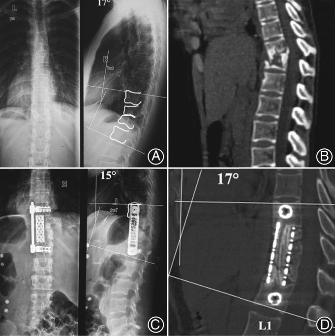
Figure 1.

A 42‐year‐old woman who sustained a T 11–12 lesion with bone destruction and kyphotic deformity was treated by anterior debridement, spinal cord decompression, distraction to correct kyphosis, and titanium cage filled with morselized rib bone and allograft bone grafting in T 11–L 1. (A, B) Preoperative radiological images are shown. (C) Postoperative roentgenographs show that the kyphosis angle was corrected by 2°. (D) Graft fusion and a 2° loss of correction in local kyphosis angle were seen 2 years postoperatively.
