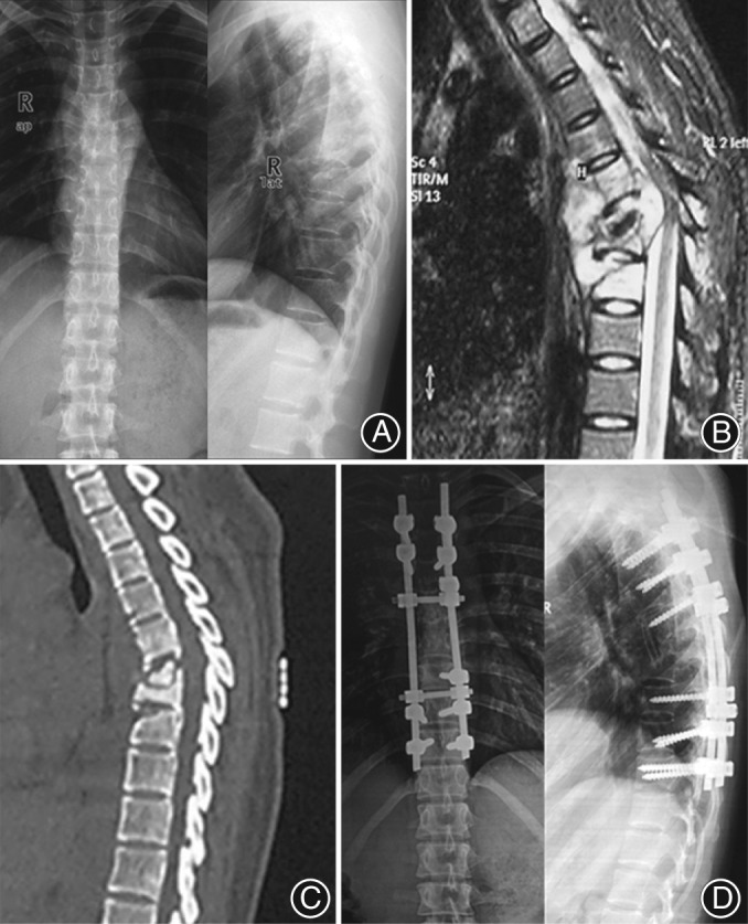
Figure 4.

A 52‐year‐old female with T 5–8 vertebrae involvement and paraplegia, who underwent a single‐stage posterior instrumentation combined with anterior debridement and rib bone grafting. (A–C) Preoperative radiography, computed tomography (CT) scan, and magnetic resonance images are shown. (D) Radiography demonstrated that satisfactory graft union was achieved 3 years postoperatively. ASIA Grade was B preoperatively and was E at the final follow‐up.
