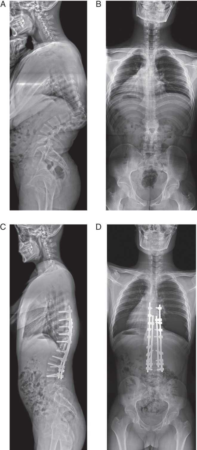
Figure 1.

Radiographs of a 21‐year‐old man with marked kyphosis secondary to thoracolumbar anomalies. (A) There are hemivertebrae at T11 and T12, and a wedged vertebra at L1. They converge anteriorly, thus separating the posterior column. (B) A transverse plane of a vertebra (L2) is clearly visible in this standing posteroanterior film. After double VCRs at T11 and T12 and use of satellite rods, (C) kyphosis was satisfactorily corrected and (D) balance well maintained.
