
Figure 2.
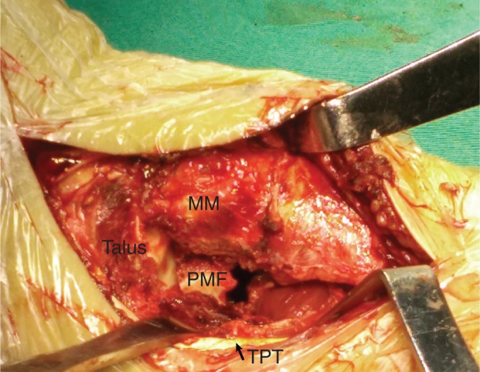
A retractor is used to pull the tibialis posterior tendon and flexor digitorum longus posteriorly. The posterior malleolar fragment and the distal tibial articular surface are clearly visualized. MM, medial malleolus; PMF, posterior malleolar fragment; TPT, tibialis posterior tendon.