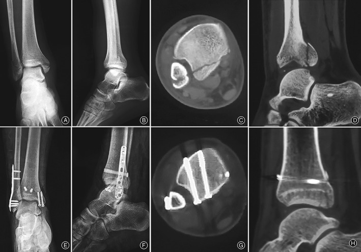
Figure 5.

The figure showed the right ankle of a 48‐year‐old female caused by a ground‐level fall, AO type 44B3. Preoperative mortise (A), lateral (B) radiographs and axial (C), sagittal (D) computed tomography (CT) scans of the trimalleolar fracture in the posteromedial group. Postoperative mortise (E) and lateral (F) radiographs and axial (G), sagittal (H) CT scans of the patient showed anatomic reduction with anteroposterior screws fixation.
