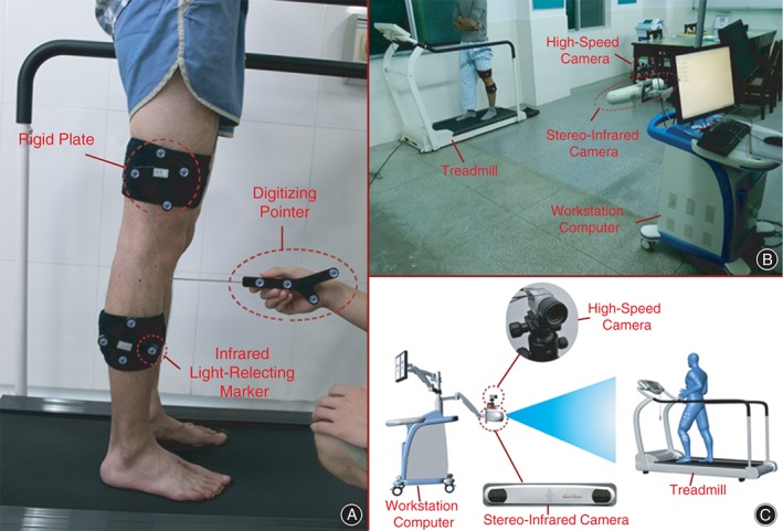
Figure 2.

(A) Identification of surface markers. Two rigid plates, each composed of four infrared light‐reflecting markers, are attached to each subject's thighs and shanks with bandages. A digitizing pointer composed of four infrared light‐reflecting markers is used to identify femoral and tibial landmarks. (B) The instrument for knee kinematics analysis. The 3‐D trajectories of the rigid plates during activities are tracked by an integrated two‐head stereo‐infrared camera at a frequency of 60 Hz. An integrated synchronous high‐speed camera is used to divide the walking cycle and to record video of the activity. A workstation computer with customized software performs supplementary real‐time calculations. The entire system is integrated on a mobile cart (1.1 m × 0.6 m × 0.4 m). Each subject was asked to walk on a horizontal treadmill at a speed of 3 km/h. (C) Schematic representation of the instrument for knee kinematics analysis.
