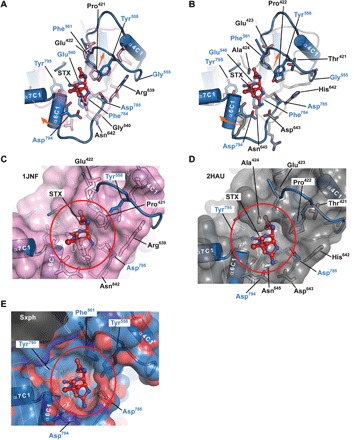
Fig. 4. Sxph STX-binding site and transferrin proto-pocket.

(A and B) Superposition of the Sxph (marine) STX-binding site with (A) Fe3+-bound rabbit serum transferrin (PDB: 1JNF) (41) (pink) and (B) apo-human serum transferrin (PDB: 2HAU) (32) (gray). STX (red) is shown as sticks. Select residues are shown. Blue labels indicate Sxph residues. Orange arrows indicate changes between transferrin and Sxph. (C to E) Transferrin proto-pocket and Sxph STX-binding pocket comparisons. (C) to (E) show apo-transferrin (pink), transferrin (gray), and Sxph (marine) surfaces, respectively. In (C) and (D), labels indicate Sxph residues that break through the transferrin surface. Red circle highlights the STX-binding site. STX is shown as space filling. Sxph surface is colored by atom type, where red and blue denote oxygen and nitrogen, respectively.
