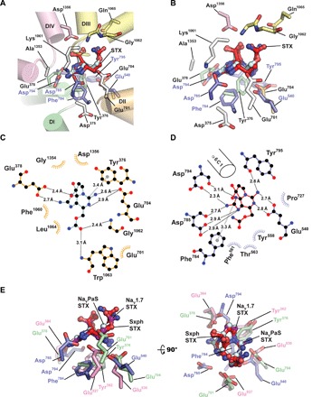
Fig. 5. Sxph and NaVs share STX recognition strategies.

(A) NaVPaS:STX (PDB: 6a91) (29) and Sxph:STX STX-binding site superposition. NaVPaS is shown as a cartoon viewed from the central channel cavity. Pore domains are colored as follows: DI, green; DII, orange; DIII, yellow; and DIV, pink. STX coordinating and selectivity filter “DEKA” motif (white) residues are shown as sticks. Sxph STX-binding site side chains are blue. STX from Sxph:STX (red) and NaVPaS:STX (cyan) are superposed. (B) Closeup view of the STX-binding sites from (A). (C) Diagram of the NaVPaS:STX interactions. (D) Diagram of the Sxph:STX interactions. (E) Comparison of common STX interactions for Sxph (blue), NaVPaS (green), and NaV1.7 (magenta). STX from the Sxph:STX complex (red), NaVPaS:STX complex (cyan), and NaV1.7:STX complex (violet) are indicated. (C) and (D) were generated using LIGPLOT (67) and a 3.35-Å cutoff. Hydrogen bonding networks (black dashed lines) and cation-π interactions (gray dashed lines) are indicated. (D) is the same as Fig. 3C.
