Figure 1.

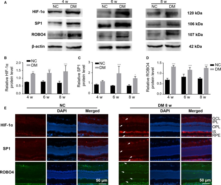
The expression levels of HIF‐1α, SP1 and ROBO4 were increased in the diabetic retina. A‐D, Western blots of HIF‐1α, SP1 and ROBO4 expression in the retinas of diabetic rats after 4, 6 and 8 weeks. β‐Actin was used as a loading control. Bars, mean ± SDs. *P < 0.05; **P < 0.01; ***P < 0.001 versus the respective negative control group (n = 6). E, Immunofluorescent staining in normal or diabetic retinas and nuclear staining by DAPI (magnification: 20×). HIF‐1α (red), SP1 (red) and ROBO4 (green) showed enhanced fluorescence (arrows) in NFL, GCL, IPL, OPL, IS and RPE layers in the diabetic group compared with normal rats. NFL: nerve fibre layer, GCL: ganglion cell layer, IPL: inner plexiform layer, OPL: outer plexiform layer, IS: inner segment, RPE: retinal pigment epithelium
