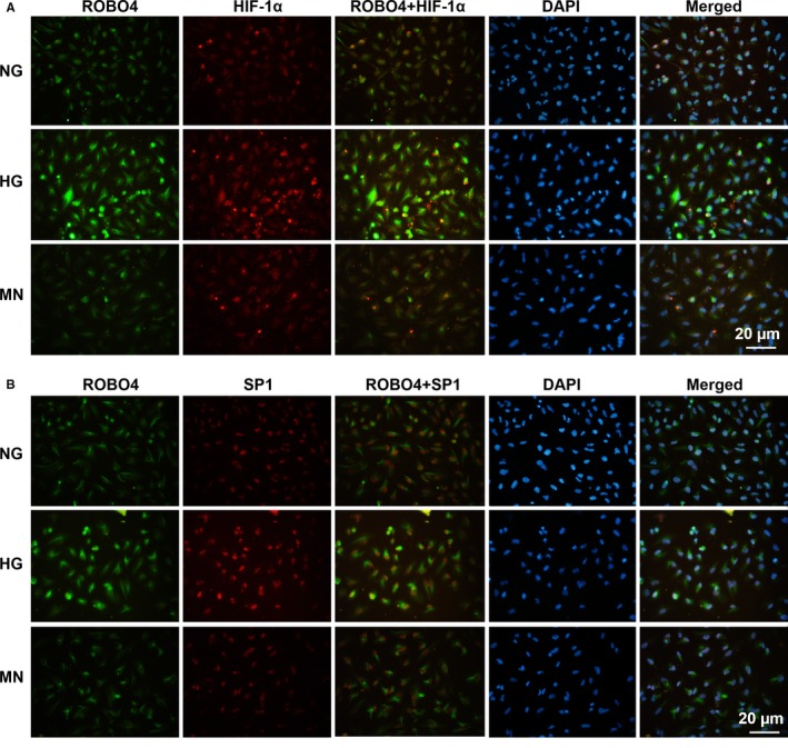
Figure 3.

Abundance and localization of HIF‐1α, SP1 and ROBO4 in ARPE‐19 cells under hyperglycaemic conditions. Dual immunostaining of HIF‐1α (red) and ROBO4 (green) A or SP1 (red) and ROBO4 (green) B, merged with DAPI (blue) in RPE cells under NG, HG and MN
