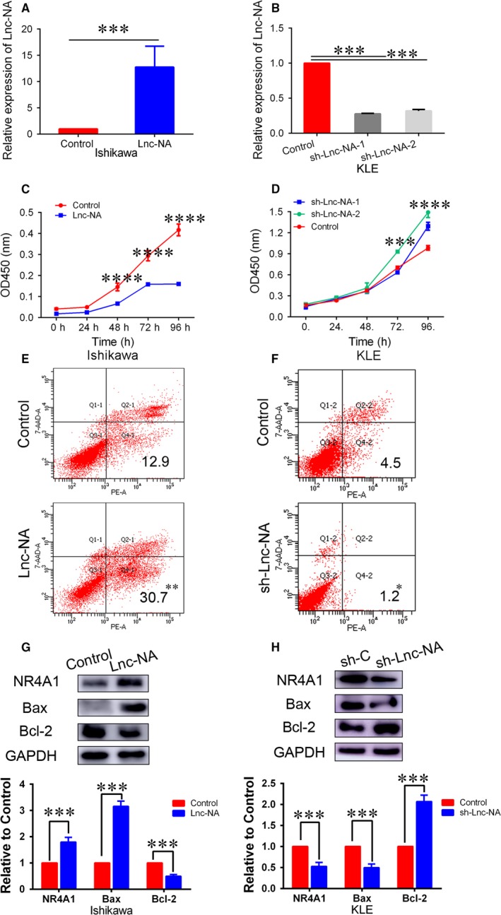
Figure 2.

Lnc‐NA inhibited cell proliferation and promoted cell apoptosis in Endometrioid endometrial carcinoma cells. (A) qRT‐PCR analysis of Lnc‐NA expression levels after Lnc‐NA overexpression in Ishikawa cells. (B) qRT‐PCR analysis of Lnc‐NA expression levels after Lnc‐NA knockdown in KLE cells. (C) CCK‐8 assays were performed to determine cell proliferation activities after transfection for 0, 24, 48, 72, and 96 h. The data show that compared with the negative groups, Lnc‐NA overexpression significantly decreased cell proliferation. (D) CCK‐8 assays were performed to determine cell proliferation activities after transfection for 0, 24, 48, 72, and 96 h. The data show that, compared with the negative groups, Lnc‐NA knockdown significantly increased cell proliferation. (E) FACS analysis was used to determine the per cent of Annexin V+/7‐AAD‐ (apoptotic) cells in Ishikawa cells transfected with the Lnc‐NA construct or negative control. (F) FACS analysis was used to determine the per cent of Annexin V+/7‐AAD‐ (apoptotic) cells in KLE cells transfected with the sh‐Lnc‐NA construct or negative control. (G) nuclear receptor subfamily 4 group A member 1 (NR4A1), Bcl2 and Bax expression levels in Ishikawa cells after transfection with the Lnc‐NA construct or negative control were determined by Western blot. (H) NR4A1, Bcl2, and Bax expression levels in Ishikawa cells after transfection with the sh‐Lnc‐NA construct or negative control were determined by Western blot. All data are shown as the means ± SD, n = 3. Significant differences between groups are indicated as *P < 0.05, **P < 0.01, ***P < 0.001, and ****P < 0.0001
