
Figure 2.
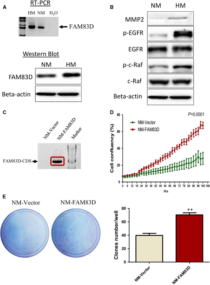
High expression of FAM83D in highly metastatic cells and the role of FAM83D in the proliferation of ovarian cancer cells. (A) Detection of FAM83D mRNA and protein levels from HM and NM cells using reverse‐transcript PCR (RT‐PCR) and Western blotting. (B) Western blotting analysis of the MMP2 and EGFR‐related pathway. (C) RT‐PCR showed the high expression of FAM83D in the stable cell line NM‐FAM83D. The FAM83D/pcDNA‐ or pcDNA (control vector)‐transfected cells were transferred into NM cells and used G418 for selection for one month. (D) The proliferation of NM cells after overexpression of FAM83D was measured using the IncuCyte® S3 Live‐Cell Analysis System for 102 h. (E) The ability for single cancer cell tumorigenesis was evaluated through a colony formation assay. The data are presented as means ± SEM; **P < 0.01 against the NM‐vector control. HM, highly metastatic; NM, non‐metastatic; RT‐PCR, reverse transcriptase–polymerase chain reaction; MMP2, matrix metalloproteinase‐2; EGFR, epidermal growth factor receptor; SEM, standard error of mean