Figure 4.

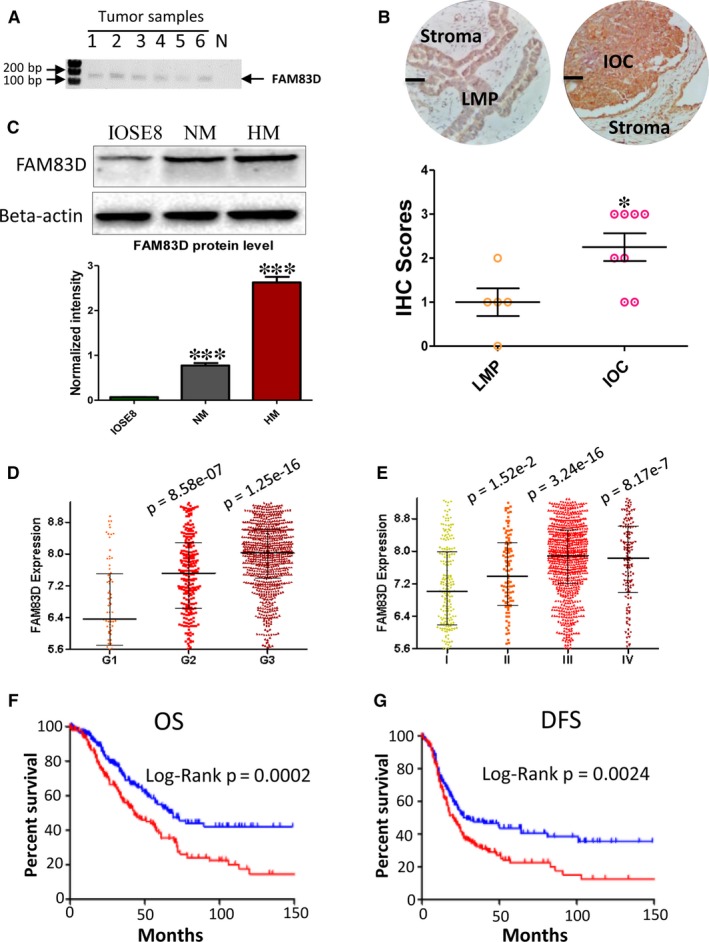
Strong FAM83D expression in IOC patient samples and high expression of FMA83D are positively associated with prognosis. (A) RT‐PCR was performed to detect the mRNA expression of FAM83D in formalin‐fixed paraffin‐embedded tumour tissues. (B) IHC staining of FAM83D in IOC and LMP samples. Upper: The representative staining of FAM83D in IOC and LMP samples (Scale bar = 50 μm). Lower: The IHC scores of IOC and LMP samples IOC (n = 8) and LMP (n = 5) (*P = 0.024). (C) The protein levels of FAM83D in immortalized ovary cell (IOSE8) and ovarian cancer cells (NM and HM) were detected using Western blotting. (D) The expression level of FAM83D in different grades of tumour tissue. The Mann‐Whitney test was performed to test other groups against the grade 1 group. (E) The expression level of FAM83D in different stages of metastasis in tumour tissues. The Mann‐Whitney test was performed to test other groups against the stage 1 group. (F) The overall survival curve of patients with high expression of FAM83D (upper 25%, n = 202) and low expression of FAM83D (lower 25%, n = 203). (G) The disease‐free survival curve of a patient with high expression of FAM83D (upper 25%, n = 176) and low expression of FAm83D (lower 25%, n = 177). The log‐rank test was performed to compare patient survival curves between two groups. IOC, invasive epithelial ovarian cancer; RT‐PCR, reverse transcriptase–polymerase chain reaction
