Figure 7.

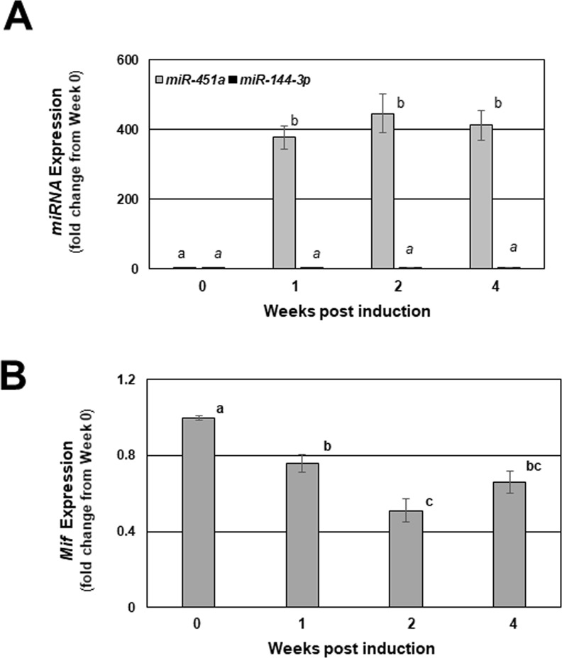
Mature miR-144-3p, miR-451a, and Mif transcript expression in endometriotic-like lesions from mice with experimentally induced endometriosis. Endometriosis was induced in wild-type mice using donor tissue from miR-144-3p/miR-451a null mice as described under “Materials and Methods” and mice were sacrificed at 1, 2 and 4 weeks post-induction (week 0 = time of induction). Endometriotic lesion-like structures were removed from each mouse, carefully minimizing underlying peritoneum contamination and total RNA was isolated. Murine miR-144-3p and miR-451a (A) were evaluated by qRT-PCR. miR-451a confirmed target transcript Mif was also evaluated by qRT-PCR in the same lesion tissues at each of the time points (B). Data are displayed as the mean ± SEM. Different letters indicate statistically significant differences among the means as determined by one-way ANOVA followed by post-hoc analysis (N = 6 per group; P < 0.05). Time 0 represents assessments made on tissue fragments (eventual lesions) that were harvested from donors but not transferred to recipient mice and served as the baseline for expression of the indicated miRNAs and their indicated targets.
