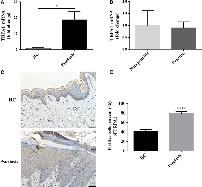
Figure 1.

TRPA1 expression is increased in skin lesions from patients with psoriasis. (A) RT‐qPCR analysis of TRPA1 expression in lesional skin from patients with psoriasis (n = 13) and normal skin from HC (n = 8). (B) TRPA1 mRNA expression in lesional skin from pruritic (n = 9) and non‐pruritic (n = 4) psoriasis patients. (C) Representative immunohistochemical images of TRPA1 expression in lesional skin from psoriasis patients and normal skin (Scale bar = 50 μm). (D) Percents of positive‐stained cells of TRPA1 in epidermis from psoriatic psoriasis (n = 13) and HC (n = 8). Results are normalized to GAPDH expression. Values were shown as mean ± SEM. (*P < 0.05, ****P < 0.0001, Student’s T test). HC, healthy controls; TRPA1, Transient receptor potential ankyrin 1
