
Figure 3.
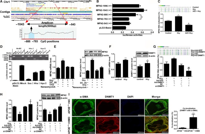
Hcy inhibited MFN2 transcriptional activities via DNMT1 in VSMCs. A, Analysis of MFN2 gene location, amplicon size and CpG islands in the amplicon. Methylation profile of CpG sites for MFN2 gene. B, The luciferase assay of activities of MFN2 promoter region (−1996/+1, −1043/+1, −885/+1, −783/+1 and −543/+1). **P < 0.01, compared to MFN (−783/+1) group. C, MassARRAY Quantitative Methylation was performed to determine methylation status at the MFN2 promoter in VSMCs. Gray circles indicate not analysed methylation values owing to CpGs with high‐ or low‐mass Dalton peaks falling outside the conservative window of reliable detection for the EpiTYPER software, the colour of the circles represents the per cent of methylation in each CpG site. D, The proximal promoter region (−1403 to +1) of MFN2 was digested with McrBC, XhoI, HhaI, HpaI and HpaII to confirm the methylation status, then Dual‐Luciferase reporter assay was performed to ascertain the activity of the MFN2 proximal promoter differentially methylated with SssI, XhoI, HhaI, HpaI and HpaII methylase in VSMCs transfected with luciferase reporter constructs. E, MFN2 expressions in VSMCs were detected by qRT‐PCR and Western blot with the co‐treatment of Hcy and DNMT1, DNMT3a and DNMT3b specific inhibitor (DC05, TFD and nanaomycin A). F, DNMT1 expression was detected by qRT‐PCR and Western blot after VSMCs were incubated with 100 μmol/L Hcy. G and H, Quantification of DNA methylation and the expression of MFN2 in VSMCs after overexpression or knock‐down of DNMT1. I, Representative immunofluorescence images of DNMT1 (red) and VSMCs marker (α‐SMA, green) in APOE−/− mice, and nuclei were stained with DAPI (blue). Yellow indicated presence of both red and green staining, scale bar, 50mm. Quantification of the average DNMT1 intensity and co‐localization with α‐SMA was done, respectively. The experiment was performed in triplicate, and the representative images are shown. *P < 0.05, **P < 0.01, compared to the APOE−/− +NC or Hcy+Ad‐GFP group, #P < 0.05, ##P < 0.01, compared to the Hcy+NC group