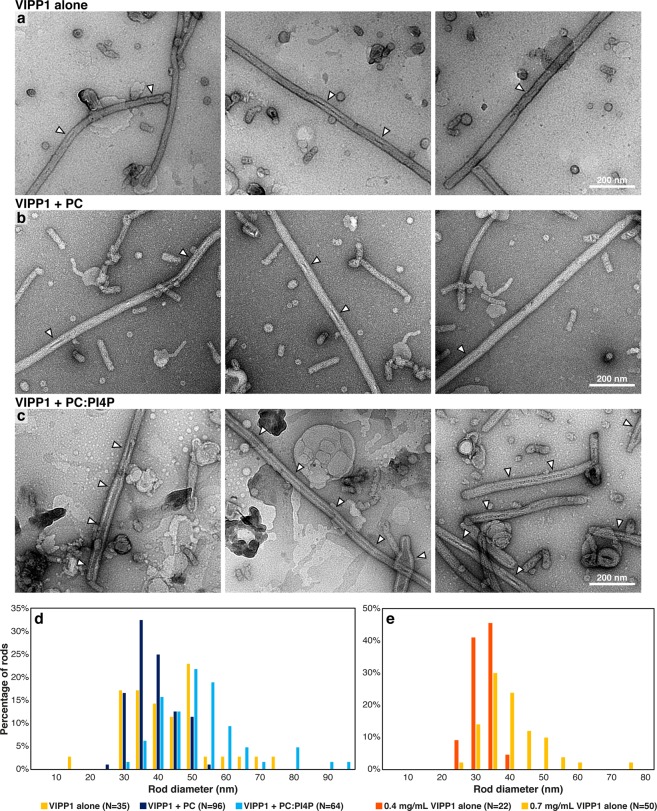
Figure 2.
VIPP1 forms large rods that can contain lipids. Negative stain electron micrographs of (a) VIPP1 alone, (b) VIPP1 incubated with PC liposomes and (c) VIPP1 incubated with PC:PI4P (95:5) liposomes. The same VIPP1 preparation (0.4 mg/mL, Prep #4, see methods) was used for all conditions, and this sample was also used for the “VIPP1 + PC:PI4P” cryo-ET in Figs. 3 and 4 (see methods). Encapsulated distinctly stained material inside the VIPP1 rods is indicated with arrowheads. (d) Distribution of rod diameters for the three conditions in (a–c). (e) Distribution of rod diameters for VIPP1 alone for two different VIPP1 preparations at concentrations of 0.4 mg/mL and 0.7 mg/mL (Preps #2 and #1, respectively, see methods). The 0.4 mg/mL sample was also used for the “VIPP1 alone” cryo-ET in Figs. 3 and 4.

