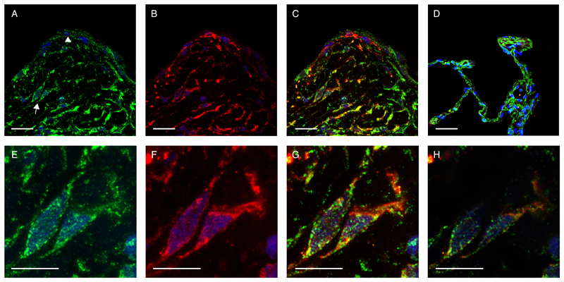
Fig. 4. ATF4 co-localises with a-SMA-positive myofibroblasts within IPF fibrotic foci.
Immunofluoresence single staining for ATF4 (A, green), α-smooth muscle actin (B, α-SMA, red) and overlay of ATF4 and α-SMA (C, yellow) in a representative IPF fibrotic focus. (A) Arrow indicates myofibroblasts within the fibrotic focus and arrowhead points to the hyperplastic epithelium. Overlay of ATF4 and α-SMA in non-IPF lung tissue (D). (E-G) Corresponding high magnification images of myofibroblasts within the fibrotic focus (indicated by arrow in A) for ATF4(E), α-SMA(F) and overlay of ATF4 and α-SMA(G). (H) Mid-level non-composite confocal overlay image indicating nuclear localisation of ATF4 (green) in an α-SMA positive myofibroblast cell (<0.5 µm). All images counter-stained with DAPI (blue). Scale A-D = 50 µm, E-H = 25 µm, N=3 patients with IPF, N=2 control subjects and representative images shown.

