Fig. 7.
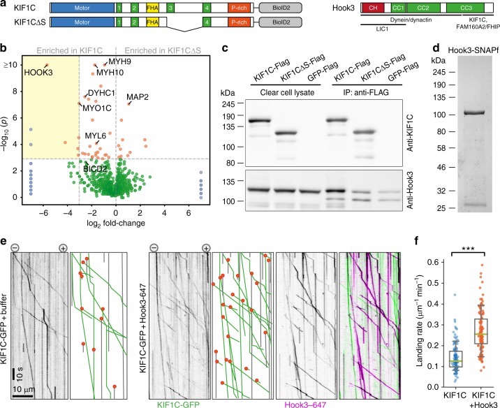
Hook3 activates KIF1C. a Schematic primary structure of constructs used for BioID: full-length KIF1C and KIF1C∆S. Hook3 is also shown together with regions of confirmed interactors. b Volcano plot shows ratio of proteins enriched in KIF1C∆S relative to full-length KIF1C relative to p-value of Fisher’s exact test. Proteins in blue were only present in one of the samples. Proteins labelled in orange are significantly enriched across the repeats. The yellow box includes all significant hits that are at least 10-fold enriched in full-length KIF1C sample. n = 3 independent experiments, one of which included three replicates. See Supplementary Data 1. c Co-immunoprecipitation of Hook3-GFP with KIF1C-Flag, KIF1C∆S-Flag and GFP-Flag from HEK cell lysates. Uncropped gels are provided in Source Data file. d Coomassie-stained SDS-PAGE of purified Hook3-SNAPf. The contamination at ~27 kDa is TEV protease used to cleave the protein off the purification tag. e Representative kymographs from single-molecule experiments of KIF1C-GFP alone or in the presence of Hook3 labelled with Alexa647. Motility and landing events indicated as in c. f Quantification of absolute frequency of KIF1C-GFP motors landing on microtubules, n = 116 and 131 MTs, respectively, pooled from three independent experiments. ***p < 0.0005 (t-test). Data are provided in Source Data file

