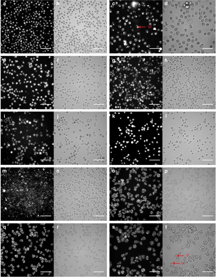
Fig. 2.
Specific feature detection of infected A. castellanii Neff by high-content screening. All cells are stained with SYBR Green. The scale bars indicate 100 µm. a SYBR Green channel for APMV at 24 h pi. b Brightfield channel for APMV at 24 h pi. c, d higher magnification of APMV at 24 h pi (scale bar indicates 50 µm). Bright spots representing the viral factory (vf) are well differentiated from the nucleus (n) and the vacuoles (v). e, f Marseillevirus T19 at 10 h pi. g, h Pandoravirus massiliensis at 18 h pi. i, j Tupanvirus Deep Ocean at 24 h pi. k, l Pacmanvirus at 6 h pi. m, n Cedratvirus at 20 h pi. o, p Faustovirus E12 at 48 h pi. q, r Orpheovirus IHUMI - LCC2 at 48 h pi. s, t negative control A. castellanii Neff at 48 h pi at high-magnification (scale bar indicates 50 µm)

