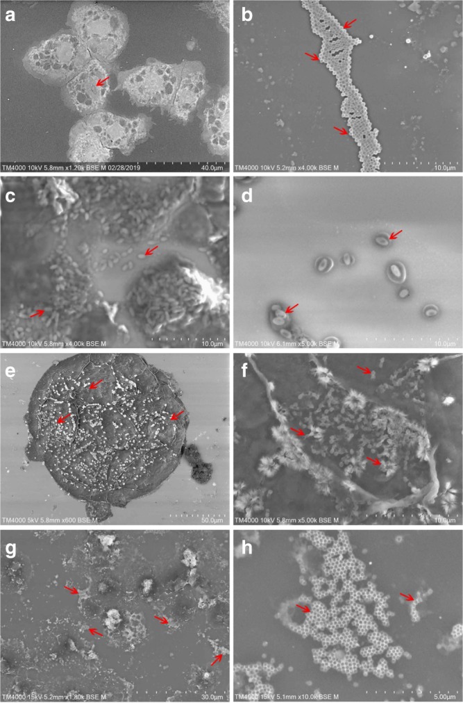
Fig. 4.

SEM images of culture supernatants showing some of the isolated giant viruses. These photos are generated from our samples using the TM4000 Plus microscope. a Uninfected A. castellanii Neff (red arrow indicates nucleus). b Mimivirus particles showing a typical ~ 650 nm capsid (red arrows). c, d Pandoravirus particles with their characteristic apical aspect. e A. castellanii Neff cell with Tupanvirus particles adhered to its surface (red arrows). f High-magnification image of e showing typical Tupanvirus particles with their characteristic tails (red arrows). g, h Supernatant of an infected culture showing clusters of Marseillevirus particles with a ~250 nm capsid (red arrows indicate clustered particles). Scale bar and acquisition settings are generated automatically by the SEM on the original micrographs
