Figure 4.
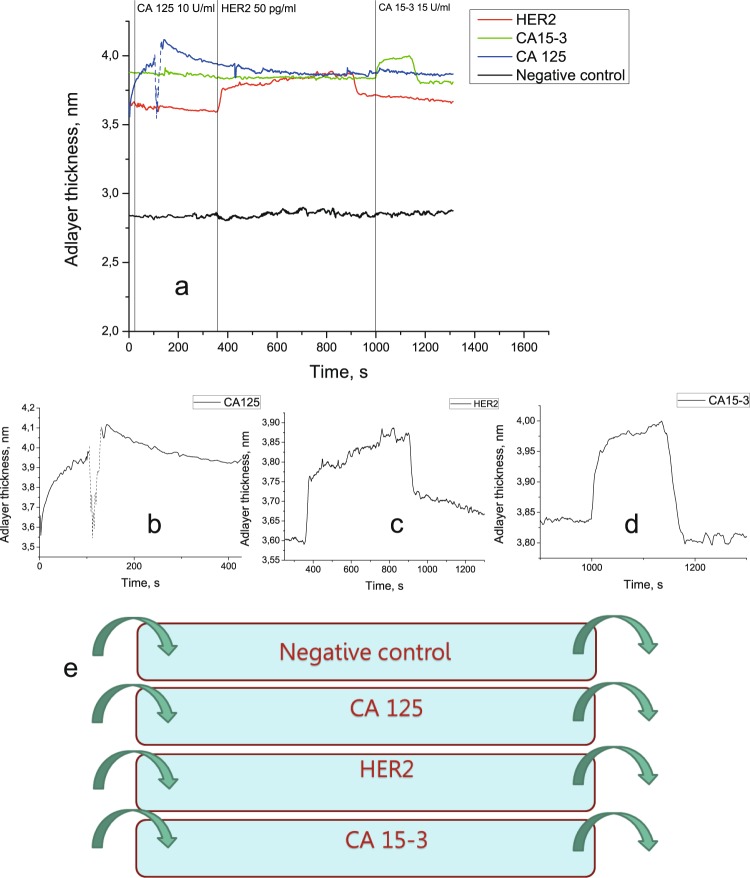
The experiment with four-channel flow cell. (a) Parallel detection of three soluble tumor markers in a four-channel flow cell containing antibodies against CA125, HER2, and CA15-3. The dip in the signal corresponding to CA125 detection (dashed line) is caused by an air bubble formation inside of the flow cell. (b) A signal corresponding to CA125 detection. (c) A signal corresponding to HER2 detection. (d) A signal corresponding to CA15-3 detection. (e) A schematic diagram of the four-channel flow cell used in the experiment.

