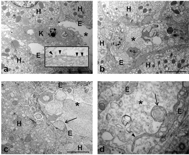
Figure 1.
Transmission electron micrographs of human liver of chronically HCV-infected patient. (a) Representative image of a preserved liver sinusoidal endothelium (E) and neighboring hepatocytes (H); a Kupffer cell (K) is visible into the sinusoidal lumen (asterisk). The inset shows intact cytoplasmic processes of sinusoidal endothelial cells with typical fenestrae (arrowheads). (b) The image show the sinusoidal lumen (asterisk) crowded by the presence of inflammatory cells and material originating from damaged cells. (c,d) Microvilli (arrowhead) extending from the surface of endothelial cells (E) and the presence of engulfed material (arrows) inside the cytoplasm are visible. Scale bars: a, b, c = 5 μm; d = 1.2 μm.

