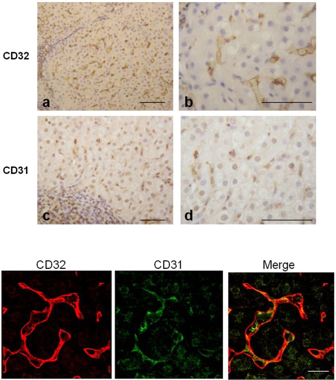
Figure 4.

Expression of CD32 and CD31 in liver from HCV-infected. (Upper panel) Immunohistochemical analysis showed positive immunoreactions of both CD32 and CD31. (Lower panel) Confocal microscope images reveals a diversity in the localization of the two markers (CD32 is expressed at the cell surface; CD31 is present inside the cytoplasm) as demonstrated by the separated colors in the merged of the fluorescence signals. Scale bars: Upper panel a,c = 100 μm; b,d = 70 μm; Lower panel 25 μm.
