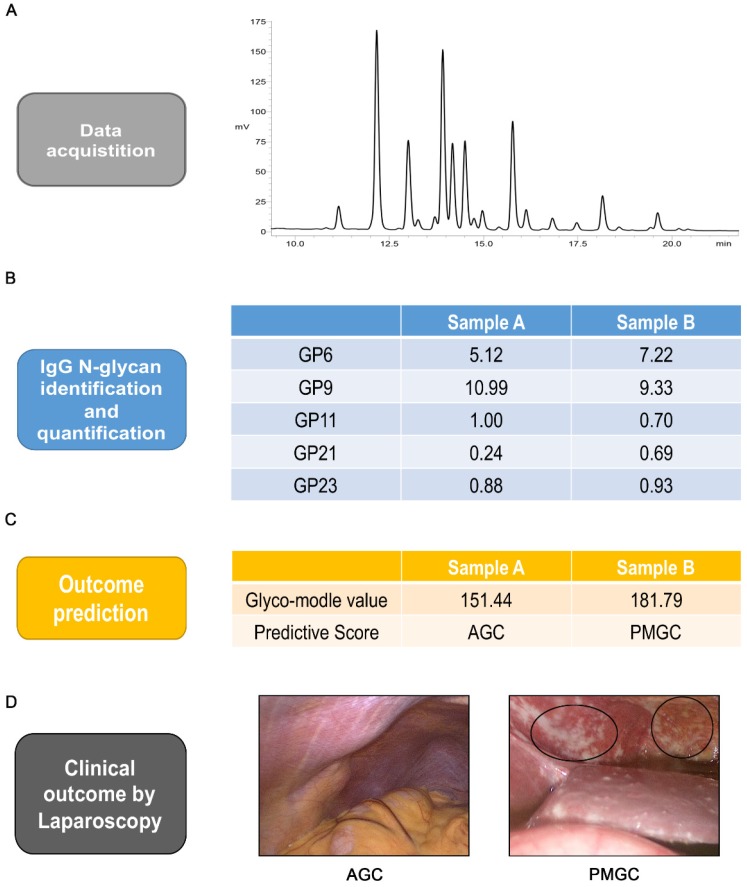
Figure 4.
Analysis workflow of prediction. Typical base peak of the serum specimen in positive ion mode(A). Identification and quantification of the five IgG glycan of GP6, GP9, GP11, GP21 and GP23 (B). Logistic regression predictive score and outcome prediction of the two samples(C). Typical images of abdominal cavity by staging laparoscopy (D). The circled parts are typical peritoneal metastasis.

