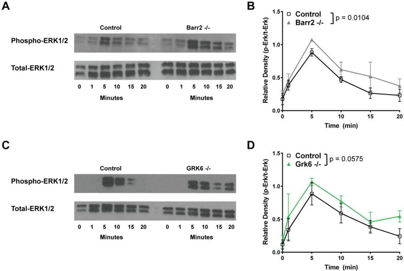
FIGURE 5.
Barr2−/− pro-inflammatory macrophages show increased ERK1/2 phosphorylation after activation by chemerin. Control versus Barr2−/− (A, B) or GRK6−/− (C, D), C57BL/6J peritoneal macrophages were either left unstimulated (0 minutes) or stimulated with 6.25 nM chemerin ex vivo for 1, 5, 10, 15, or 20 minutes. Total protein was normalized using a BCA assay. Western blot analysis was performed on cell lysates probed using phospho-p44/42 MAPK (ERK1/2) (Thr202/Tyr204) (Phospho-ERK1/2) and p44/42 MAPK (ERK1/2) (Total-ERK1/2). Representative blots (A, C) of 3 (Barr2−/−) or 4 (GRK6−/−) independent experiments are shown. Quantification shown in (B, D) was done by densitometry using ImageJ software and is shown as a mean ratio of relative density ± SEM (Barr2−/− n=3; GRK6−/− n=4).

