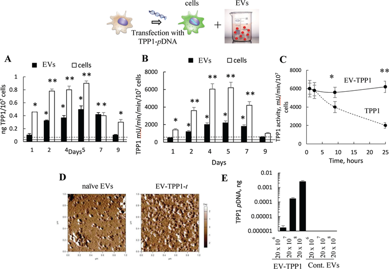
Figure 1.
Characterization of EVs isolated from TPP1-transfected macrophages (EV-TPP1-t). TPP1-transfected macrophages and their EVs display elevated levels of A) TPP1 protein and B) enzymatic activity between second and seventh day post-transfection. IC21 macrophages were transfected with TPP1-encoding pDNA (2 μg mL−1 pDNA with GenePorter 3K for 4 h), and (A) TPP1 protein expression and (B) TPP1 enzymatic activity were assessed in the parental cells (white bars) and EVs (black bars) using (A) ELISA and (B) a TPP1 substrate, AF-AMC (400 μM), respectively. (A) TPP1 protein or (B) activity levels in nontransfected cells (dashed line) or EVs secreted by them (solid line) were also recorded. For EVs’ activity, the levels are normalized to the number of cells used to isolate these EVs. C) Increased stability of TPP1 in EVs (solid line) compared to EV-free TPP1 (dashed line) upon incubation with pronase protease from Streptomyces Greseus (4 × 10⁻⁵ M). D) Round morphology of EVs released by empty-transfected macrophages and TPP1-transfected macrophages. E) Quantitative PCR analysis indicated a significant amount of TPP1-encoding pDNA incorporated in EVs from pretransfected macrophages. (A–C) Statistical significance *p < 0.05, or **p < 0.005 compared to TPP1 levels in nontransfected cells or (A,B) EVs, or (E) EV-free TPP1.

