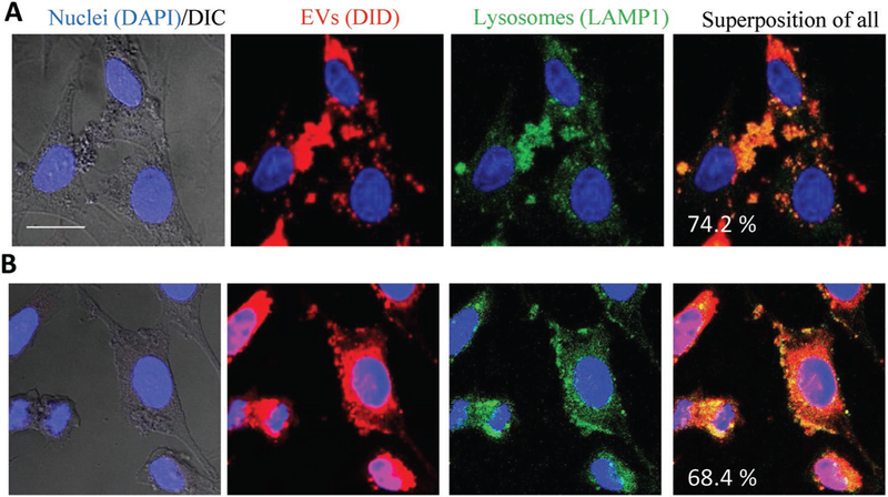
Figure 4.

EVs target lysosomal compartments in PC12 neuronal cells. The cells were incubated with DID-labeled EVs (1010 particles/ml) for A) 1 h or B) 4 h, and then stained with FITC-LAMP1 antibodies for lysosomes and DAPI for nuclei. Colocalization of EVs (red) and lysosomes (green) is manifested in yellow. The bar: 20 μm.
