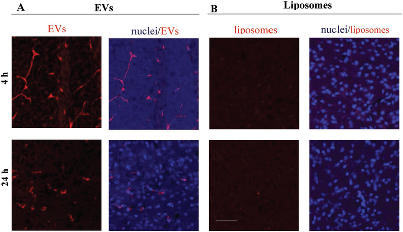
Figure 6.

Brain distribution of DiD-labeled A) EVs and B) liposomes in knock-out LINCL mice. Confocal images showed a strong fluorescence in different brain areas in case of DiD-EV, and low, if any, fluorescence in case of DiD-liposomes. 1 week old knock-out mice were injected i.p. with DiD-labeled (A) EVs or (B) liposomes (1010 particles/100 μL/mouse). Animals were sacrificed 4 h or 24 h after injections and perfused. Brain slides were processed and examined by confocal microscopy. Nuclei were stained by DAPI (blue). The bar: 50 μm. EVs were isolated from macrophages concomitant media, and labeled with fluorescent dye, DiD (red). Liposomes were prepared and labeled with DiD (red).
