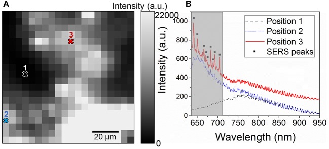
Figure 4.
(A) 100 × 100 μm2 map of the integrated intensity in the 640–710 nm performed on a LFM revealed with gold NPs with a 5 μm spatial resolution and (B) Raman spectra acquired at three different positions of the map showing the spectrum of glass (Position 1), a region of the fingerprint with gold NPs (Position 2), and a region with SERS enhancement (Position 3, black squares). The gray region indicates the integrated intensity range.

