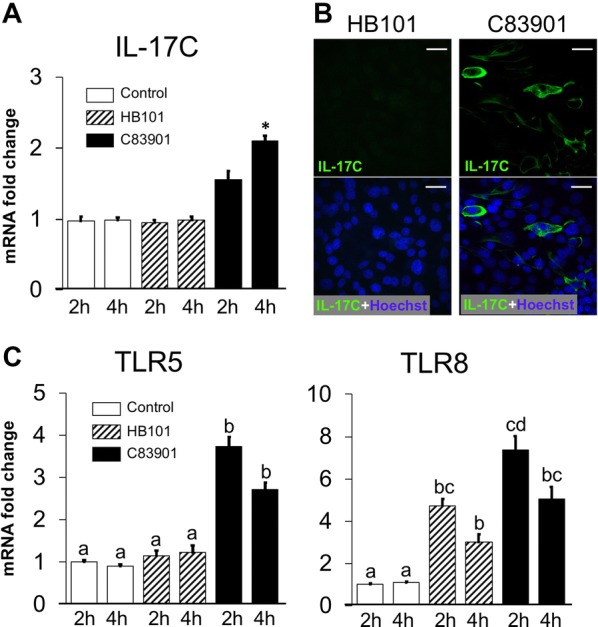
Figure 2.

Induction of IL-17C and TLR-5, -8 in the IPEC-J2 cells after F4+ETEC infection. IPEC-J2 monolayers were inoculated with F4+ ETEC (C83901), non-pathogenic E. coli strain HB101 at MOI 100 or PBS for 1 h and further incubated for another 1 h, 3 h or 23 h, respectively. The mRNA expression of IL-17C (A) and TLR5 and TLR8 (C) in the IPEC-J2 monolayers was assessed by qPCR. The mRNA expression level was normalized to the reference genes and then to the control group of 2 h treatment. Data are presented as the mean ± SD (n = 3 per group), different letters indicate significant differences between groups (p < 0.05). B IPEC-J2 monolayers were inoculated with the different bacterial strains at MOI 100 for 1 h and then further incubated for 23 h. The cells were stained with anti-IL-17C (FITC, green). The nuclei were counterstained with Hoechst and representative confocal images are shown (n = 3). Scale bar = 25 μm.
